Calm, dignified
experience

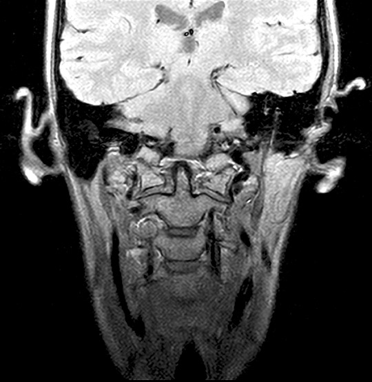
Craniocervical junction MRI scan
A craniocervical junction MRI scan can give detailed images of the junction between the bone at the base of the skull and the top two bones in the cervical spine (in the neck).
MRI Cranio-Cervical Junction Scan (AI Enhanced)
Medserena’s Craniocervical Junction MRI scan
From £1,495.00
MRI scan of the Craniocervical Junction and Cervical Spine; non-invasive procedure to help diagnose medical conditions relating to the junction between the base of the skull and the cervical
spine, price includes:
- Open and Upright MRI scan
- variPOSETM flexion, extension and rotational views
- 90 minutes appointment
- Radiologist findings report
- Images provided via secure link to your email address at the end of the scan and available to NHS trusts via IEP on request
- Complimentary refreshments
Please wear metal free clothing and if possible, avoid wearing any jewellery. Alternatively, Medserena can provide you with a gown to change into for your scan. Scroll down for more Craniocervical Junction MRI scan information, variations and options.
Many Scans Available within 48 hours
Craniocervical Junction MRI scan variation and pricing
| CCJ & Cervical Spine + variPOSE |
From £1,495.00 |
| CCJ & Brain & Cervical Spine + variPOSE |
From £1,765.00 |
| CCJ & Cervical Spine + variPOSE & Thoracic |
From £1,845.00 |
| CCJ & Cervical Spine + variPOSE & Lumbar + variPOSE |
From £2,085.00 |
| CCJ & Cervical Spine + variPOSE & Thoracic & Lumbar + variPOSE |
From £2,435.00 |
| CCJ and Brain and Cervical spine + variPOSE and Thoracic and Lumbar + variPOSE |
From £2,480.00 |
Superior healthcare service with every Private MRI scan
Little or no
waiting time
Largest MRI scan centres
Premium
refreshments
Watch TV while
scanning
Medical report included
About Craniocervical Junction MRI scans
The craniocervical junction (CCJ) is where the skull joins the spine and includes the occipital bone at the base of the skull and the first two bones of the spine called the axis and atlas bones. The atlas allows for the movement of your head. The opening at the base of the occipital bone is called the foramen magnum and it protects the brain stem, important nerves, and blood vessels.
This type of MRI scan is a useful imaging investigation for people suffering with neck pain, often accompanied by headaches at the back of the neck. This type of pain usually gets worse with movements such as coughing or bending forwards.
Medserena’s craniocervical junction MRI scan is one of the company’s clinical specialities and offers an upright scan that can be carried out with the patient in a comfortable position to allow full assessment of the foramen magnum and cerebellar tonsils (these are structures in the brain located at the base of the cerebellar hemispheres, two halves of the cerebellum (the part of the brain which control movements and balance). The scan takes around one and a half hours and the cerebellar tonsils can be assessed under the natural weight of gravity. This may show a more accurate degree of hind brain herniation (also known as coning), where part of the brain is squeezed through the foramen magnum. This can cause a range of neurological symptoms.
The scan is carried out in an open scanner and a variety of different views are taken using the company’s unique variPOSE sequence, for example showing the head turned to the right, ahead and then left, giving a rotational dynamic study. The neck is also scanned in flexion (bending), extension and neutral positions. Static measurements are then taken and compared to standardised normal levels. Further measurements are taken in flexion and extension to assess the degree of movement that person has. The alar ligaments are one of the key areas assessed to see if they are lax or damaged.
The big advantage of an open craniocervical junction MRI scan over a conventional flatbed MRI tunnel scan is that the upright spine can be viewed in a neutral upright position whilst weight bearing under the natural load of gravity. This gives a dynamic picture of what is happening in the CCJ and upper spine when it is subjected to normal everyday pressures picking up problems that might have been missed or underestimated on a conventional flatbed MRI scanner.
A craniocervical junction MRI scan is particularly useful for patients with whiplash type symptoms and those with the hypermobile connective tissue disease Ehlers-Danlos syndromes. Many ED patients complain their head feels too heavy and say that they are asymmetrical.
What conditions can a craniocervical MRI scan detect?
- Craniocervical instability (CCI): Craniocervical Junction MRI scans can determine if craniocervical instability is present. This is a condition which can cause constant headaches and a heavy head feeling. It is structural instability at the junction of where the skull meets the spine which makes it prone to excessive movement. This can lead to ligaments becoming stretched, weakened, or ruptured. It is common in patients with the inherited hypermobility condition Ehlers-Danlos syndromes, a group of rare conditions which affect connective tissue. Some people develop CCI after a whiplash injury from a car crash.
- Fusion of the atlas (C1) and occipital bone: The Atlanto-occipital joint is where the very top of the spine connects to the hole in the base of the skull. These bones can fuse, causing compression of the main arteries in the neck and the spinal cord. Symptoms include headaches, neck pain, dizziness, trouble swallowing and it can be difficult to distinguish these from those of Chiari malformation (see below).
- Rheumatoid Arthritis: This is an inflammatory form of arthritis which can cause joints to swell and become inflamed, ultimately damaging them. This can lead to acquired CCJ instability.
- Paget disease: This is a condition where the body develops abnormal bone turnover and an area of softened and enlarged bone grows. It is common in over 50s and can also lead to instability and weakness in the neck.
- Basilar Invagination: This is where the very top of the neck is pushed too far up into the foramen magnum. The neck will appear shortened and can lead to compression of vital structures.
- Ehlers-Danlos Syndromes: These are a group of rare inherited conditions that affect the connective tissues in ligaments, tendons, skin, blood vessels, bones and internal organs, causing loose unstable joints prone to dislocation, and fragile stretchy skin. The condition can often be misdiagnosed as ME/chronic fatigue.
- Chiari malformation: Type 1 Chiari malformation is where the lower part of the brain pushes down onto the spinal canal, and sometimes requires surgery. This is a condition that is present from birth but not usually discovered until adulthood. Some people have no symptoms and the Chiari malformation is only diagnosed when they have an MRI scan. These can be assessed more accurately in an upright scanner rather than lying flat in a tunnel scanner.

Benefits of having variPOSE during
your open, upright MRI scan
The variPOSE sequence includes comprehensive MRI scans of the spine or CCJ whilst sitting in neutral, flexion (bending), extended or rotational positions, showing both the axial, sagittal, and coronal anatomical views (similar to the planes of the body).
Other benefits of a Medserena craniocervical junction MRI scan
Open MRI scanners are a stress-free alternative to using a conventional enclosed tunnel MRI scanner, providing comfort and reassurance for people who suffer from anxiety or claustrophobia. Sitting upright is more comfortable for patients and the open front means patients can see a friend or relative or watch television throughout as distraction.
Open MRI scans can also accommodate larger/ heavier patients who might have difficulty fitting comfortably into a conventional tunnel scanner, as they can take weights of up to 35 stone (226kg). However, suitability will depend on the patient’s build and the area of anatomy that needs to be scanned.
FAQs
The Upright MRI is truly open. There are no tunnels, no narrow tubes. The system is particularly quiet, the examination is comfortable and does not trigger feelings of being in a confined space. This means that the Upright MRI is particularly tolerated by patients who suffer from “claustrophobia”.
Because the system offers you an unrestricted view, you can watch TV or see DVD movies on a large screen during the scan. Wearing headphones – as with other MRI systems – is usually not necessary.
According to the current state of knowledge, there is no danger to the patient’s health as magnetic resonance imaging only uses magnetic fields and radio waves.
Metallic foreign bodies within the patient, such as fixed dental prosthesis, artificial joints or metal plates after treatment for a fracture do not usually pose any danger. However, it is important to clarify that the implants you use are MRI-compatible before the examination.
MRI (Magnetic Resonance Imaging) utilises a large magnet, radio waves and a computer to form images of your body. It is non-invasive, painless and does not use any ionising radiation.
Our truly open MRI can scan you in different positions. Through the utilisation of a specially designed MRI system we can offer weight-bearing scans – sitting or standing. The design of the system allows the patient to be positioned in different postures (e.g. flexion or extension) so that the patient may be examined in the position where they experience pain. The reason to do this is that some pathologies are underestimated or even not seen in a conventional supine MRI scan. The technique has value in many applications: e.g. spine, knees, hips, ankles. This has been proven in scientific studies and documented in peer reviewed publications.
In addition, it offers the possibility of performing an MRI scan on patients who could not otherwise tolerate the examination. This may include the claustrophobic patient, who benefits from the truly open nature of the equipment, and the severely kyphotic patient or emphysema sufferer who simply cannot lie down. It can also facilitate scanning of large patients who struggle to fit conventional ‘bore’ MRI scanners.
Of course, we have a comfortable waiting area but if you want them to stay in the scan room with you, they will also need to fill out a safety questionnaire.There is enough space for a companion to sit in a chair close to the scanner to offer support. This particularly beneficial when examining a teenager.
This depends above all on which part of the body needs to be examined. In the Upright MRI, special examinations can be carried out in various body positions. The entire scan generally takes between 30 and 45 minutes. However, since you have the opportunity to watch TV or DVD, this time will go by much quicker.
Eat and drink normally and, unless your doctor tells you otherwise, please continue taking medications as normal. If you have any special needs (e.g. wheelchair access) please inform us when making the appointment.
Your appointment confirmation; referral letter/form; Medical Insurance details if applicable. We accept all major debit/credit cards.
We will provide a gown/clothing for you to wear when you are scanned. If you prefer to wear your own, please ensure that you wear or bring clothing without any metal fasteners, zips or under-wiring as these cannot be worn in the scan room. The changing room can be locked for safe storage of your possessions.
You will be able to walk into the scanner. It has no tunnel or bore. You will be able to hear us and talk with us during your scan if necessary-and we will be able to see you at all times. Due to its open nature, you will even be able to watch TV or a DVD whilst having the scan. Depending on which part of you is being scanned, you may be asked to sit or stand, and assume different postures (for example bending forward.) The radiographer may place a receiver “coil” around the relevant area of your body. You will need to remain very still while the acquisition is done in order to prevent blurring of the images. You will hear some tapping from the scanner but in general it is much quieter than many other MRI scanners.
You will not feel anything while having the scan. There is no pain or unusual feeling of any type and you will experience no after effects.
YES. There are some things that can prevent you from having an MRI scan. You will be asked to complete a safety questionnaire on arrival at the Centre which will cover the contra-indications-but if you are making an appointment and any of the factors below affect you, please discuss this with us in advance as it may save you a wasted trip.
Contra-indications can include:
- Pacemaker
- IUDs
- Surgical clips
- Pregnancy
- Metal fragments in the body
- Metal pins/plates/screws
- Joint replacements
- Metal objects in eyes
- Cochlear implants
- IVC filters
- Metal heart valves
- Penile implants
It is also important to tell us if you have any tattoos or piercings.
Watches, jewellery, coins, keys, cigarette lighters, penknives, credit cards. piercings, hairgrips, wigs, nicotine patches, and hearing aids must be removed.
Your scan will be reported by a Consultant Radiologist. It will normally be available in a couple of days unless needed urgently. The images and report will be sent to your referring practitioner. If you have a follow up appointment, please make us aware of the details so we can ensure the report and images are available in time.