Calm, dignified
experience

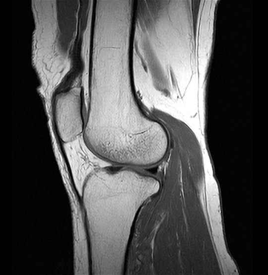
Knee MRI scan
A knee MRI scan can reveal the inner workings of this weight-bearing joint, discovering the causes of pain, stiffness and swelling.
MRI Knee Sagittal Scan (AI Enhanced)
Medserena Knee MRI scan
From £430.00
MRI scan of the knee; non-invasive procedure to help diagnose medical conditions relating to the tendons, muscles, cartilage, ligaments and bones of the knee joint, price includes:
- Open and Upright MRI scan
- 45 minutes appointment
- Radiologist findings report
- Images provided via secure link to your email address at the end of the scan and available to NHS trusts via IEP on request
- Complimentary refreshments
Please wear metal free clothing and if possible, avoid wearing any jewellery. Alternatively, Medserena can provide you with a gown to change into for your scan. Choose and book your scan now, or scroll down for more knee joint MRI scan information.
Many scans available within 72 hours
Knee MRI scan variation and pricing
| Knee |
From £430.00 |
| Knees Bilateral |
From £660.00 |
Superior healthcare service with every Private MRI scan
Little or no
waiting time
Largest MRI scan centres
Premium
refreshments
Watch TV while
scanning
Medical report included
About Knee MRI scans
Knee pain is a common and debilitating problem causing pain and restricting movement – particularly as people get older. One study in the over 50s, found 47 per cent had experienced knee pain in the previous 12 months, with one third of those seeing their GP about their pain. The most common causes of knee pain are related to aging, injury, or repeated stress on the knee.
Having a knee MRI scan in the upright position can reveal the structure of the knee in a load-bearing position, meaning a more accurate assessment of the alignment of the kneecap, ligaments and so on. It can also highlight any instability of the knee.
In our scanners, the knee will be examined in the standing inclined positions, which strikes a balance between upright load-bearing and patient comfort.
Osteoarthritis, caused by ‘shock-absorbing’ cartilage wearing away between bones, is the most common cause of knee pain in older people and around 70,000 knee replacement operations are performed every year. The knee is composed of a complex network of ligaments and the inner cartilage, called meniscus, is frequently torn in sports such as football. It also weakens with age and can be torn by twisting motions. Due to its excellent soft tissue contrast a knee MRI scan can give the most complete picture of what is going on.
What problems can a knee MRI scan detect?
There are many knee conditions visible on an MRI scan, too numerous to list them all here, but they include:
- Meniscal tears: Tears in the meniscus, a c-shaped piece of cartilage between the femur (thigh bone) and tibia (shin bone), are usually caused by activities such as squatting, heavy lifting, or turning suddenly on the sports field. It can be accompanied by a popping noise, as well as pain, swelling and difficulties moving the knee and it can feel unstable.
- Anterior Cruciate Ligament/Poster Cruciate Ligament injuries: These are the main ligaments at the front and back of the knee. A sprained or strained knee ligament or muscle is usually caused by a blow to the knee or a sudden twist of the knee. Symptoms often include pain, swelling, and difficulty walking.
- Chondromalacia patellae (runner’s knee): Pain behind the patella (kneecap) when active or in certain positions, is caused by softening of cartilage in this area. Pain is triggered by walking, running, going up and down stairs, kneeling, squatting, and sitting for extended periods with knee bent. It has many causes including overuse, injury, arthritis, or inflammation of the lining of the joint.
- Patellofemoral pain syndrome: This is one of the most common knee sports injuries and refers to pain at the front, around and behind the kneecap. It causes a dull, aching, pain which is usually worse with activities where you bend your knee, or if you sit with your knees bent for a long time.
- Inflammation of the tendons may result from overuse of a tendon during certain activities such as running, jumping, or cycling. Tendonitis of the patellar tendon is called jumper's knee. This often occurs with sports, such as basketball, where the force of hitting the ground after a jump strains the tendon.
- Tibial Plateau fractures: Tibial plateau fractures are complex injuries of the knee. The tibial plateau is one of the most critical load-bearing areas in the human body. Tibial plateau fractures are typically caused by a strong force on the lower leg and are commonly seen in road traffic accidents, sports accidents with a high velocity such as skiing, horse riding, and certain water sports
Other benefits of a Medserena Knee MRI Scan
Open MRI scanners are a stress-free alternative to using a conventional enclosed tunnel MRI scanner, providing comfort and reassurance for people who suffer from anxiety or claustrophobia. Patients are scanned upright and tilted back and the open front means patients can see a friend or relative and watch television throughout as a distraction.
Open MRI scans can also accommodate larger/ heavier patients who might have difficulty fitting comfortably into a conventional tunnel scanner, as they can take weights of up to 35 stone (226kg). However, suitability will depend on the patient’s build and the area of the anatomy to be scanned.
Available to self-pay clients, clients with private health insurance and NHS patients where prior funding has been agreed by a clinical commissioning group.
FAQs
The Upright MRI is truly open. There are no tunnels, no narrow tubes. The system is particularly quiet, the examination is comfortable and does not trigger feelings of being in a confined space. This means that the Upright MRI is particularly tolerated by patients who suffer from “claustrophobia”.
Because the system offers you an unrestricted view, you can watch TV or see DVD movies on a large screen during the scan. Wearing headphones – as with other MRI systems – is usually not necessary.
According to the current state of knowledge, there is no danger to the patient’s health as magnetic resonance imaging only uses magnetic fields and radio waves.
Metallic foreign bodies within the patient, such as fixed dental prosthesis, artificial joints or metal plates after treatment for a fracture do not usually pose any danger. However, it is important to clarify that the implants you use are MRI-compatible before the examination.
MRI (Magnetic Resonance Imaging) utilises a large magnet, radio waves and a computer to form images of your body. It is non-invasive, painless and does not use any ionising radiation.
Our truly open MRI can scan you in different positions. Through the utilisation of a specially designed MRI system we can offer weight-bearing scans – sitting or standing. The design of the system allows the patient to be positioned in different postures (e.g. flexion or extension) so that the patient may be examined in the position where they experience pain. The reason to do this is that some pathologies are underestimated or even not seen in a conventional supine MRI scan. The technique has value in many applications: e.g. spine, knees, hips, ankles. This has been proven in scientific studies and documented in peer reviewed publications.
In addition, it offers the possibility of performing an MRI scan on patients who could not otherwise tolerate the examination. This may include the claustrophobic patient, who benefits from the truly open nature of the equipment, and the severely kyphotic patient or emphysema sufferer who simply cannot lie down. It can also facilitate scanning of large patients who struggle to fit conventional ‘bore’ MRI scanners.
Of course, we have a comfortable waiting area but if you want them to stay in the scan room with you, they will also need to fill out a safety questionnaire.There is enough space for a companion to sit in a chair close to the scanner to offer support. This particularly beneficial when examining a teenager.
This depends above all on which part of the body needs to be examined. In the Upright MRI, special examinations can be carried out in various body positions. The entire scan generally takes between 30 and 45 minutes. However, since you have the opportunity to watch TV or DVD, this time will go by much quicker.
Eat and drink normally and, unless your doctor tells you otherwise, please continue taking medications as normal. If you have any special needs (e.g. wheelchair access) please inform us when making the appointment.
Your appointment confirmation; referral letter/form; Medical Insurance details if applicable. We accept all major debit/credit cards.
We will provide a gown/clothing for you to wear when you are scanned. If you prefer to wear your own, please ensure that you wear or bring clothing without any metal fasteners, zips or under-wiring as these cannot be worn in the scan room. The changing room can be locked for safe storage of your possessions.
You will be able to walk into the scanner. It has no tunnel or bore. You will be able to hear us and talk with us during your scan if necessary-and we will be able to see you at all times. Due to its open nature, you will even be able to watch TV or a DVD whilst having the scan. Depending on which part of you is being scanned, you may be asked to sit or stand, and assume different postures (for example bending forward.) The radiographer may place a receiver “coil” around the relevant area of your body. You will need to remain very still while the acquisition is done in order to prevent blurring of the images. You will hear some tapping from the scanner but in general it is much quieter than many other MRI scanners.
You will not feel anything while having the scan. There is no pain or unusual feeling of any type and you will experience no after effects.
YES. There are some things that can prevent you from having an MRI scan. You will be asked to complete a safety questionnaire on arrival at the Centre which will cover the contra-indications-but if you are making an appointment and any of the factors below affect you, please discuss this with us in advance as it may save you a wasted trip.
Contra-indications can include:
- Pacemaker
- IUDs
- Surgical clips
- Pregnancy
- Metal fragments in the body
- Metal pins/plates/screws
- Joint replacements
- Metal objects in eyes
- Cochlear implants
- IVC filters
- Metal heart valves
- Penile implants
It is also important to tell us if you have any tattoos or piercings.
Watches, jewellery, coins, keys, cigarette lighters, penknives, credit cards. piercings, hairgrips, wigs, nicotine patches, and hearing aids must be removed.
Your scan will be reported by a Consultant Radiologist. It will normally be available in a couple of days unless needed urgently. The images and report will be sent to your referring practitioner. If you have a follow up appointment, please make us aware of the details so we can ensure the report and images are available in time.