Calm, dignified
experience

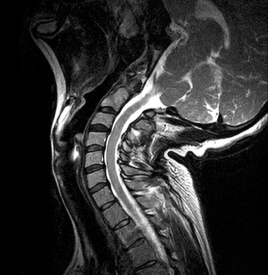
Cervical Spine MRI scan
A cervical spine MRI scan can provide answers for the causes of neck pain, stiffness and reduced range of movement.
MRI Cervical Spine VariPOSE Scan (AI Enhanced)
Medserena Cervical Spine MRI Scan
From £625.00
MRI scan of the Cervical spine; non-invasive procedure to help diagnose medical conditions relating to the neck, price includes:
- Open and Upright MRI scan
- 30 minutes appointment
- Radiologist findings report
- Images provided via secure link to your email address at the end of the scan and available to NHS trusts via IEP on request
- Complimentary refreshments
Please wear metal free clothing and if possible, avoid wearing any jewellery. Alternatively, Medserena can provide you with a gown to change into for your scan. Scroll down for more cervical spine MRI scan information, variations and options.
Many Scans Available within 48 hours
Cervical spine MRI scan variation and pricing
| Cervical spine |
From £625.00 |
| Cervical spine & Thoracic |
From £870.00 |
| Cervical spine & Lumbar |
From £870.00 |
| Cervical spine & Lumbar + standing |
From £970.00 |
| From £1,215.00 | |
| Cervical spine & Shoulder |
From £915.00 |
| Cervical spine + variPOSE |
From £725.00 |
| Cervical spine + variPOSE & Shoulder |
From £1,120.00 |
| Cervical spine + variPOSE & Thoracic |
From £1,075.00 |
| Cervical spine + variPOSE & Lumbar + variPOSE |
From £1,315.00 |
| Cervical spine + variPOSE & Lumbar + variPOSE + standing |
From £1,415.00 |
| From £1,665.00 |
Please note:
variPOSE refers to seated flexion and extension imaging. Standing is a separate lumbar spine add-on and is only included if specifically selected at the time of booking. Standing scans can be booked during the Centre’s core hours only (Monday-Friday 9am-5:30pm).
Superior healthcare service with every Private MRI scan
Little or no
waiting time
Largest MRI scan centres
Premium
refreshments
Watch TV while
scanning
Medical report included
About Cervical Spine MRI Scans
It examines the seven bones called vertebrae that make up the cervical segment of the spine between the base of the skull and the upper back. These vertebrae are stacked on top of each other to form the spinal column.
The spinal column supports your head and protects the spinal cord. This is the main structure which links the network of nerves throughout your body.
MRI allows this column to be viewed in 3D, as well as the ligaments, tendons, nerves, muscles, and blood supply that surround it.
Neck pain is common as the cervical region of the spine has to support the heavy weight of the head (which can weigh as much as 11 lbs) and allow for a flexible range of motion to turn the head.
Symptoms include pain, muscle spasms, pins and needles, weakness, restricted movement, and nerve pain, as well as pain and weakness and loss of feeling in the arms. Patients often report pain in their shoulders.
Neck pain can occur as a result of injury, wear and tear diseases such as osteoarthritis, nerve compression, muscle spasm, instability, infections, and cervical spondylitis. Pain is classified as chronic when it has a duration of 12 weeks or more. Chronic neck pain often presents as widespread increased sensitivity to pain to touch and in both passive and active movements in neck and shoulder area.
Advantages of a Medserena Cervical Spine MRI Scan
The big advantage of an open cervical spine MRI Scan over a conventional flatbed MRI tunnel scan is that the upright spine can be viewed in a neutral upright position. It can also be viewed from various angles including sitting, leaning forward, and stretching back, using a variPOSE sequence of MRI scans, so it is seen when weight bearing under the natural load of gravity.
This gives a dynamic picture of what is happening in the spine when it is subjected to normal everyday pressures such as sitting and standing, picking up problems that might have been missed or underestimated on a conventional flatbed MRI scanner.
In a seated position it may be possible to appreciate the true dimensions of disc bulges and how far they may slip forward or backwards in alignment compared to being laid flat, as well as potentially a truer picture of how far the canal width is reduced.
The seated and open nature of this scanner can also make it more comfortable for patients whose pain is brought on by laying on their back to be scanned. It can also be easier to scan patients whose spine is scoliotic (curved sideways), or kyphotic (an excessive outward curve of the spine resulting in an abnormal rounding of the upper back).
Conditions a cervical spine MRI scan can detect
These include:
- Cervical spondylosis: Commonly referred to as arthritis of the neck, this condition comes from wear and tear of muscles and bones in the neck region, leading to thinning of cartilage in the spinal discs. The resulting pain and stiffness in the neck and shoulders can cause headaches at the back of the head. This can also include herniated discs in the neck.
- Craniocervical instability (CCI): Cervical Spine MRI scans can determine if craniocervical instability is present. This is a condition which can cause constant headaches and a heavy head feeling. It is a structural instability at the junction where the skull meets the spine which makes it prone to excessive movement. This can lead to ligaments becoming stretched, weakened or ruptured. It is common in patients with the inherited hypermobility condition Ehlers Danlos syndrome, a disease which affects connective tissue. Some people develop CCI after a whiplash injury from a car crash.
- Internal damage to the spinal cord: This can happen due to neurodegenerative conditions such as multiple sclerosis, where the myelin sheath cover of nerves is damaged.
- Inflammatory diseases: Axial spondyloathritis (also known as AxSpA) is a form of inflammatory arthritis that can cause pain and stiffness in the neck and other areas of the spine, as well as other joints. The inflammation is visible on an MRI scan but not on an x-ray. If AxSpA develops into ankylosing spondylitis, where vertebrae fuse together, causing pain and restricting movement, this will be visible on an x-ray.
- Malignant tumours: These will be visible on a cervical spine MRI scan.
- Cervical myelopathy: Spinal cord nerve damage is caused by disc prolapse in the spine or bony spurs forming which compress the spinal cord. It causes permanent damage if not treated. The symptoms usually develop slowly. Because of the lack of pain, there may be an interval of years between the onset of disease and first treatment. Early symptoms of this condition are numb, clumsy, painful hands and reduced fine motor skills.

Benefits of having variPOSE during
your open, upright MRI scan
The variPOSE sequence includes comprehensive MRI scans of the spine or CCJ whilst sitting in neutral, flexion (bending), extended or rotational positions, showing both the axial, sagittal, and coronal anatomical views (similar to the planes of the body).
Other benefits of a Medserena cervical spine MRI scan
Open MRI scanners are a stress-free alternative to using a conventional enclosed tunnel MRI scanner, providing comfort and reassurance for people who suffer from anxiety or claustrophobia. Sitting upright is more comfortable for patients and the open front means patients can see a friend or relative or watch television throughout as distraction.
Open MRI scans can also accommodate larger/ heavier patients who might have difficulty fitting comfortably into a conventional tunnel scanner, as they can take weights of up to 31.5 stone (200kg). However, suitability will depend on the patient’s build and the area of anatomy that needs to be scanned.
- Book a Medserena cervical spine MRI scan direct in London or Manchester
FAQs
The Upright MRI is truly open. There are no tunnels, no narrow tubes. The system is particularly quiet, the examination is comfortable and does not trigger feelings of being in a confined space. This means that the Upright MRI is particularly tolerated by patients who suffer from “claustrophobia”.
Because the system offers you an unrestricted view, you can watch TV or see DVD movies on a large screen during the scan. Wearing headphones – as with other MRI systems – is usually not necessary.
According to the current state of knowledge, there is no danger to the patient’s health as magnetic resonance imaging only uses magnetic fields and radio waves.
Metallic foreign bodies within the patient, such as fixed dental prosthesis, artificial joints or metal plates after treatment for a fracture do not usually pose any danger. However, it is important to clarify that the implants you use are MRI-compatible before the examination.
MRI (Magnetic Resonance Imaging) utilises a large magnet, radio waves and a computer to form images of your body. It is non-invasive, painless and does not use any ionising radiation.
Our truly open MRI can scan you in different positions. Through the utilisation of a specially designed MRI system we can offer weight-bearing scans – sitting or standing. The design of the system allows the patient to be positioned in different postures (e.g. flexion or extension) so that the patient may be examined in the position where they experience pain. The reason to do this is that some pathologies are underestimated or even not seen in a conventional supine MRI scan. The technique has value in many applications: e.g. spine, knees, hips, ankles. This has been proven in scientific studies and documented in peer reviewed publications.
In addition, it offers the possibility of performing an MRI scan on patients who could not otherwise tolerate the examination. This may include the claustrophobic patient, who benefits from the truly open nature of the equipment, and the severely kyphotic patient or emphysema sufferer who simply cannot lie down. It can also facilitate scanning of large patients who struggle to fit conventional ‘bore’ MRI scanners.
Of course, we have a comfortable waiting area but if you want them to stay in the scan room with you, they will also need to fill out a safety questionnaire.There is enough space for a companion to sit in a chair close to the scanner to offer support. This particularly beneficial when examining a teenager.
This depends above all on which part of the body needs to be examined. In the Upright MRI, special examinations can be carried out in various body positions. The entire scan generally takes between 30 and 45 minutes. However, since you have the opportunity to watch TV or DVD, this time will go by much quicker.
Eat and drink normally and, unless your doctor tells you otherwise, please continue taking medications as normal. If you have any special needs (e.g. wheelchair access) please inform us when making the appointment.
Your appointment confirmation; referral letter/form; Medical Insurance details if applicable. We accept all major debit/credit cards.
We will provide a gown/clothing for you to wear when you are scanned. If you prefer to wear your own, please ensure that you wear or bring clothing without any metal fasteners, zips or under-wiring as these cannot be worn in the scan room. The changing room can be locked for safe storage of your possessions.
You will be able to walk into the scanner. It has no tunnel or bore. You will be able to hear us and talk with us during your scan if necessary-and we will be able to see you at all times. Due to its open nature, you will even be able to watch TV or a DVD whilst having the scan. Depending on which part of you is being scanned, you may be asked to sit or stand, and assume different postures (for example bending forward.) The radiographer may place a receiver “coil” around the relevant area of your body. You will need to remain very still while the acquisition is done in order to prevent blurring of the images. You will hear some tapping from the scanner but in general it is much quieter than many other MRI scanners.
You will not feel anything while having the scan. There is no pain or unusual feeling of any type and you will experience no after effects.
YES. There are some things that can prevent you from having an MRI scan. You will be asked to complete a safety questionnaire on arrival at the Centre which will cover the contra-indications-but if you are making an appointment and any of the factors below affect you, please discuss this with us in advance as it may save you a wasted trip.
Contra-indications can include:
- Pacemaker
- IUDs
- Surgical clips
- Pregnancy
- Metal fragments in the body
- Metal pins/plates/screws
- Joint replacements
- Metal objects in eyes
- Cochlear implants
- IVC filters
- Metal heart valves
- Penile implants
It is also important to tell us if you have any tattoos or piercings.
Watches, jewellery, coins, keys, cigarette lighters, penknives, credit cards. piercings, hairgrips, wigs, nicotine patches, and hearing aids must be removed.
Your scan will be reported by a Consultant Radiologist. It will normally be available in 5 working days. The images and report will be sent to your referring practitioner. If you have a follow up appointment, please make us aware of the details so we can ensure the report and images are available in time.