Calm, dignified
experience

Sacroiliac joints MRI scan
A sacroiliac joint MRI scan shows the sacroiliac joints that join the central sacrum bone to the ilium bones of the pelvis on each side.
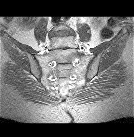
MRI Sacroiliac joint coronal view Scan (AI Enhanced)
Medserena Sacroiliac Joint MRI Scan
From £520.00
MRI scan of the Sacroiliac Joint; non-invasive procedure to help diagnose medical conditions relating to the base of the spine, price includes:
- Open and Upright MRI scan
- 30 minutes appointment
- Radiologist findings report
- Images provided via secure link to your email address at the end of the scan and available to NHS trusts via IEP on request
- Complimentary refreshments
Please wear metal free clothing and if possible, avoid wearing any jewellery. Alternatively, Medserena can provide you with a gown to change into for your scan. Scroll down for more Sacroiliac Joint MRI scan information.
Many Scans Available within 48 hours
Sacroiliac joints MRI scan variation and pricing
| Sacroiliac joints |
From £520.00 |
| Lumbar Spine & Sacroiliac joints |
From £860.00 |
Superior healthcare service with every Private MRI scan
Little or no
waiting time
Largest MRI scan centres
Premium
refreshments
Watch TV while
scanning
Medical report included
About Sacroiliac Joint MRI Scans
The sacroiliac joints support the spine and bear the weight of the body. They are supported by strong ligaments extending around the back of the pelvis. The glutes and piriformis muscle also help to reinforce the joint. An MRI scan may show up inflammation which may not be visible with other imaging techniques such as CT scans and x-ray.
Up to 30 per cent of people with mechanical lower back pain have inflammation of the sacroiliac joints, also referred to as sacroiliitis or sacroiliac joint syndrome.
The big advantage of an open sacroiliac MRI scan over a conventional flatbed MRI tunnel scan is that the upright spine can be viewed in a neutral upright position whilst sitting, so it is seen when weight bearing under the natural load of gravity.
This gives a true picture of what is happening in the SI joints when it is subjected to normal everyday pressures when sitting, picking up problems that might have been missed or underestimated on a conventional flatbed MRI scanner.
The seated and open design of this scanner is also usually more comfortable for patients whose pain is brought on by lying on their back to be scanned. It can also be easier to scan patients whose spine is scoliotic (curved sideways), or kyphotic (an excessive outward curve of the spine resulting in an abnormal rounding of the upper back).
What conditions can a sacroiliac joint MRI scan detect?
These include:
- Sacroiliac joint dysfunction (SJD)/sacroiliitis: A sacroiliac joint MRI scan can reveal early stages of inflammation in the joints that might not be visible on an x-ray. SJD is caused by too much or too little movement of the joint, as they typically only move a small amount. Symptoms of SJD include sciatic type pain in the buttocks or back of the thigh or a stiff lower back and is sometimes accompanied by a low-grade fever. Causes can include falls or car crash injuries, infections, osteoarthritis and injury to ligaments in pregnancy as well as inflammatory arthritis (see below).
- Axial Spondyloarthritis (or spondyloarthropathy): Axial spondyloathritis (also known as AxSpA) is a form of inflammatory rheumatic arthritis that can cause pain and stiffness in the sacroiliac joints and other areas of the spine, as well as other joints. The inflammation is visible on an MRI scan but not on an x-ray. Inflammation of the Entheses (where tendons and ligaments attach to the bone) is very common with this condition.
- The most well-known type of inflammatory arthritis is ankylosing spondylitis, which mainly affects the spine but also the sacroiliac joints. Ankylosing spondylitis symptoms include pain and stiffness that doesn’t improve with rest, but gets better with exercise, buttock area pain and lower back pain that wakes you at night. It has a much earlier onset than osteoarthritis, with symptoms beginning in people in their twenties or earlier. In the later stages of the disease new bone grows between the vertebrae so they effectively fuse together, causing pain and movement restriction.
- Psoriatic arthritis: A type of inflammatory arthritis which can cause joint damage, psoriatic arthritis is associated with the skin disease psoriasis. Psoriasis causes red, raised, scaly plaques, which can be itchy and painful. Symptoms are believed to be caused by cells in the immune system becoming overactive and speeding up the new skin cell replacement process. Skin cells are replaced in a few days instead of within 21 to 28 days. In a study in 2013, sacroiliac joint involvement was observed in 34 to 78 per cent of patients with psoriatic arthritis.
Other benefits of a Medserena sacroiliac joint MRI Scan
Open MRI scanners are a stress-free alternative to using a conventional enclosed tunnel MRI scanner, providing comfort and reassurance for people who suffer from anxiety or claustrophobia. Sitting upright is more comfortable for patients and the open front means patients can see a friend or relative or watch television throughout as distraction.
Open MRI scans can also accommodate larger/ heavier patients who might have difficulty fitting comfortably into a conventional tunnel scanner, as they can take weights of up to 35 stone (226kg). However, suitability will depend on the patient’s build and the area of anatomy that needs to be scanned.
Available to self-pay clients, clients with private health insurance and NHS patients where prior funding has been agreed by a clinical commissioning group.
FAQs
The Upright MRI is truly open. There are no tunnels, no narrow tubes. The system is particularly quiet, the examination is comfortable and does not trigger feelings of being in a confined space. This means that the Upright MRI is particularly tolerated by patients who suffer from “claustrophobia”.
Because the system offers you an unrestricted view, you can watch TV or see DVD movies on a large screen during the scan. Wearing headphones – as with other MRI systems – is usually not necessary.
According to the current state of knowledge, there is no danger to the patient’s health as magnetic resonance imaging only uses magnetic fields and radio waves.
Metallic foreign bodies within the patient, such as fixed dental prosthesis, artificial joints or metal plates after treatment for a fracture do not usually pose any danger. However, it is important to clarify that the implants you use are MRI-compatible before the examination.
MRI (Magnetic Resonance Imaging) utilises a large magnet, radio waves and a computer to form images of your body. It is non-invasive, painless and does not use any ionising radiation.
Our truly open MRI can scan you in different positions. Through the utilisation of a specially designed MRI system we can offer weight-bearing scans – sitting or standing. The design of the system allows the patient to be positioned in different postures (e.g. flexion or extension) so that the patient may be examined in the position where they experience pain. The reason to do this is that some pathologies are underestimated or even not seen in a conventional supine MRI scan. The technique has value in many applications: e.g. spine, knees, hips, ankles. This has been proven in scientific studies and documented in peer reviewed publications.
In addition, it offers the possibility of performing an MRI scan on patients who could not otherwise tolerate the examination. This may include the claustrophobic patient, who benefits from the truly open nature of the equipment, and the severely kyphotic patient or emphysema sufferer who simply cannot lie down. It can also facilitate scanning of large patients who struggle to fit conventional ‘bore’ MRI scanners.
Of course, we have a comfortable waiting area but if you want them to stay in the scan room with you, they will also need to fill out a safety questionnaire.There is enough space for a companion to sit in a chair close to the scanner to offer support. This particularly beneficial when examining a teenager.
This depends above all on which part of the body needs to be examined. In the Upright MRI, special examinations can be carried out in various body positions. The entire scan generally takes between 30 and 45 minutes. However, since you have the opportunity to watch TV or DVD, this time will go by much quicker.
Eat and drink normally and, unless your doctor tells you otherwise, please continue taking medications as normal. If you have any special needs (e.g. wheelchair access) please inform us when making the appointment.
Your appointment confirmation; referral letter/form; Medical Insurance details if applicable. We accept all major debit/credit cards.
We will provide a gown/clothing for you to wear when you are scanned. If you prefer to wear your own, please ensure that you wear or bring clothing without any metal fasteners, zips or under-wiring as these cannot be worn in the scan room. The changing room can be locked for safe storage of your possessions.
You will be able to walk into the scanner. It has no tunnel or bore. You will be able to hear us and talk with us during your scan if necessary-and we will be able to see you at all times. Due to its open nature, you will even be able to watch TV or a DVD whilst having the scan. Depending on which part of you is being scanned, you may be asked to sit or stand, and assume different postures (for example bending forward.) The radiographer may place a receiver “coil” around the relevant area of your body. You will need to remain very still while the acquisition is done in order to prevent blurring of the images. You will hear some tapping from the scanner but in general it is much quieter than many other MRI scanners.
You will not feel anything while having the scan. There is no pain or unusual feeling of any type and you will experience no after effects.
YES. There are some things that can prevent you from having an MRI scan. You will be asked to complete a safety questionnaire on arrival at the Centre which will cover the contra-indications-but if you are making an appointment and any of the factors below affect you, please discuss this with us in advance as it may save you a wasted trip.
Contra-indications can include:
- Pacemaker
- IUDs
- Surgical clips
- Pregnancy
- Metal fragments in the body
- Metal pins/plates/screws
- Joint replacements
- Metal objects in eyes
- Cochlear implants
- IVC filters
- Metal heart valves
- Penile implants
It is also important to tell us if you have any tattoos or piercings.
Watches, jewellery, coins, keys, cigarette lighters, penknives, credit cards. piercings, hairgrips, wigs, nicotine patches, and hearing aids must be removed.
Your scan will be reported by a Consultant Radiologist. It will normally be available in a couple of days unless needed urgently. The images and report will be sent to your referring practitioner. If you have a follow up appointment, please make us aware of the details so we can ensure the report and images are available in time.