Calm, dignified
experience

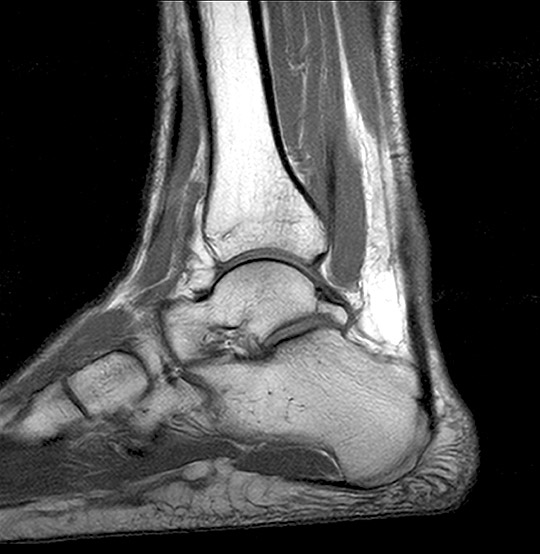
Ankle MRI scan
An ankle MRI scan can give a comprehensive overview of this joint, helping to pinpoint the causes of pain, stiffness or swelling.
MRI Ankle Scan (AI Enhanced)
Medserena Ankle MRI Scan
From £430.00
MRI scan of the ankle; non-invasive procedure to help diagnose medical conditions relating to the tendons, muscles, cartilage, ligaments and bones of the ankle joint, price includes:
- Open and Upright MRI scan
- 45 minutes appointment
- Radiologist findings report
- Images provided via secure link to your email address at the end of the scan and available to NHS trusts via IEP on request
- Complimentary refreshments
Please wear metal free clothing and if possible, avoid wearing any jewellery. Alternatively, Medserena can provide you with a gown to change into for your scan. Scroll down for more ankle
joint MRI scan information.
Many Scans Available within 48 hours
Superior healthcare service with every Private MRI scan
Little or no
waiting time
Largest MRI scan centres
Premium
refreshments
Watch TV while
scanning
Medical report included
About Ankle MRI Scans
The ankle joint is made up of tibia, fibula and talus bones, as well as the supporting ligaments, muscles, and nerves. It carries the weight of the body and can develop a range of problems, most commonly traumatic injuries of the medial and lateral malleoli (the bumps on the inside and outside of your ankle).
Ankle pain can stem from sprains and strains, to fractures and tendons or ligament problems, as well as damage to bones and cartilage due to arthritis.
An advantage of an upright ankle MRI scan over a conventional flatbed MRI tunnel scan is the ankle can be examined when partially weight-bearing (the body is angled to 20 or 40 degrees).
Other benefits of a Medserena ankle MRI scan
Open MRI scanners are a stress-free alternative to using a conventional enclosed tunnel MRI scanner, providing comfort and reassurance for people who suffer from anxiety or claustrophobia. The open front means patients can see a friend or relative or watch television throughout as a distraction.
Open MRI scans can also accommodate larger/heavier patients who might have difficulty fitting comfortably into a conventional tunnel scanner, as they can take weights of up to 31.5 stone (200kg).
Available to self-pay clients, clients with private health insurance and NHS patients where prior funding has been agreed by a clinical commissioning group.
FAQs
The Upright MRI is truly open. There are no tunnels, no narrow tubes. The system is particularly quiet, the examination is comfortable and does not trigger feelings of being in a confined space. This means that the Upright MRI is particularly tolerated by patients who suffer from “claustrophobia”.
Because the system offers you an unrestricted view, you can watch TV or see DVD movies on a large screen during the scan. Wearing headphones – as with other MRI systems – is usually not necessary.
According to the current state of knowledge, there is no danger to the patient’s health as magnetic resonance imaging only uses magnetic fields and radio waves.
Metallic foreign bodies within the patient, such as fixed dental prosthesis, artificial joints or metal plates after treatment for a fracture do not usually pose any danger. However, it is important to clarify that the implants you use are MRI-compatible before the examination.
MRI (Magnetic Resonance Imaging) utilises a large magnet, radio waves and a computer to form images of your body. It is non-invasive, painless and does not use any ionising radiation.
Our truly open MRI can scan you in different positions. Through the utilisation of a specially designed MRI system we can offer weight-bearing scans – sitting or standing. The design of the system allows the patient to be positioned in different postures (e.g. flexion or extension) so that the patient may be examined in the position where they experience pain. The reason to do this is that some pathologies are underestimated or even not seen in a conventional supine MRI scan. The technique has value in many applications: e.g. spine, knees, hips, ankles. This has been proven in scientific studies and documented in peer reviewed publications.
In addition, it offers the possibility of performing an MRI scan on patients who could not otherwise tolerate the examination. This may include the claustrophobic patient, who benefits from the truly open nature of the equipment, and the severely kyphotic patient or emphysema sufferer who simply cannot lie down. It can also facilitate scanning of large patients who struggle to fit conventional ‘bore’ MRI scanners.
Of course, we have a comfortable waiting area but if you want them to stay in the scan room with you, they will also need to fill out a safety questionnaire.There is enough space for a companion to sit in a chair close to the scanner to offer support. This particularly beneficial when examining a teenager.
This depends above all on which part of the body needs to be examined. In the Upright MRI, special examinations can be carried out in various body positions. The entire scan generally takes between 30 and 45 minutes. However, since you have the opportunity to watch TV or DVD, this time will go by much quicker.
Eat and drink normally and, unless your doctor tells you otherwise, please continue taking medications as normal. If you have any special needs (e.g. wheelchair access) please inform us when making the appointment.
Your appointment confirmation; referral letter/form; Medical Insurance details if applicable. We accept all major debit/credit cards.
We will provide a gown/clothing for you to wear when you are scanned. If you prefer to wear your own, please ensure that you wear or bring clothing without any metal fasteners, zips or under-wiring as these cannot be worn in the scan room. The changing room can be locked for safe storage of your possessions.
You will be able to walk into the scanner. It has no tunnel or bore. You will be able to hear us and talk with us during your scan if necessary-and we will be able to see you at all times. Due to its open nature, you will even be able to watch TV or a DVD whilst having the scan. Depending on which part of you is being scanned, you may be asked to sit or stand, and assume different postures (for example bending forward.) The radiographer may place a receiver “coil” around the relevant area of your body. You will need to remain very still while the acquisition is done in order to prevent blurring of the images. You will hear some tapping from the scanner but in general it is much quieter than many other MRI scanners.
You will not feel anything while having the scan. There is no pain or unusual feeling of any type and you will experience no after effects.
YES. There are some things that can prevent you from having an MRI scan. You will be asked to complete a safety questionnaire on arrival at the Centre which will cover the contra-indications-but if you are making an appointment and any of the factors below affect you, please discuss this with us in advance as it may save you a wasted trip.
Contra-indications can include:
- Pacemaker
- IUDs
- Surgical clips
- Pregnancy
- Metal fragments in the body
- Metal pins/plates/screws
- Joint replacements
- Metal objects in eyes
- Cochlear implants
- IVC filters
- Metal heart valves
- Penile implants
It is also important to tell us if you have any tattoos or piercings.
Watches, jewellery, coins, keys, cigarette lighters, penknives, credit cards. piercings, hairgrips, wigs, nicotine patches, and hearing aids must be removed.
Your scan will be reported by a Consultant Radiologist. It will normally be available in 5 working days. The images and report will be sent to your referring practitioner. If you have a follow up appointment, please make us aware of the details so we can ensure the report and images are available in time.